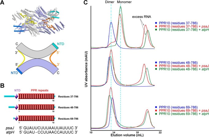
FIGURE 2.
Amino-terminal truncations lead to different dimerization states of PPR10 bound to RNA. A, upper panel: dimeric crystal structure of PPR10 in complex with psaJ RNA, two protomers of PPR10 molecules are colored light purple and gray with their NTDs colored blue and cyan, and the two bound RNA molecules are colored orange and yellow. Lower panel: a schematic illustration for dimerization state of PPR10 with psaJ RNAs. B, schematic representation of successive amino-terminal truncations on PPR10. Red boxes indicate tandem PPR repeats. The NTD is colored light purple, and loop regions are colored cyan. The sequences of two native RNA targets psaJ and atpH for PPR10 are shown below. C, SEC analyses of successive amino-terminal truncated PPR10 proteins in the absence and presence of psaJ or atpH RNA element. Dimer and monomer positions are indicated by cyan dotted lines. All structure figures were prepared with PyMOL (43). mAU, milliabsorbance units.

