FIGURE 2.
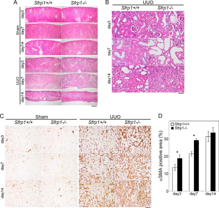
Loss of Sfrp1 exacerbates the progression of renal fibrosis after UUO. A and B, representative microscopic images of the sham and UUO kidneys in wild-type (Sfrp1+/+) and Sfrp1-deficient mice (Sfrp1−/−). These tissue sections were prepared and stained with hematoxylin and eosin. B, higher magnification is shown in A. C, αSMA immunostaining in the obstructed kidneys. D, graph shows analysis of the percentage of αSMA-positive area in the UUO kidneys. Scale bars, 500 μm (A), 50 μm (B), 100 μm (C). *, p < 0.05.

