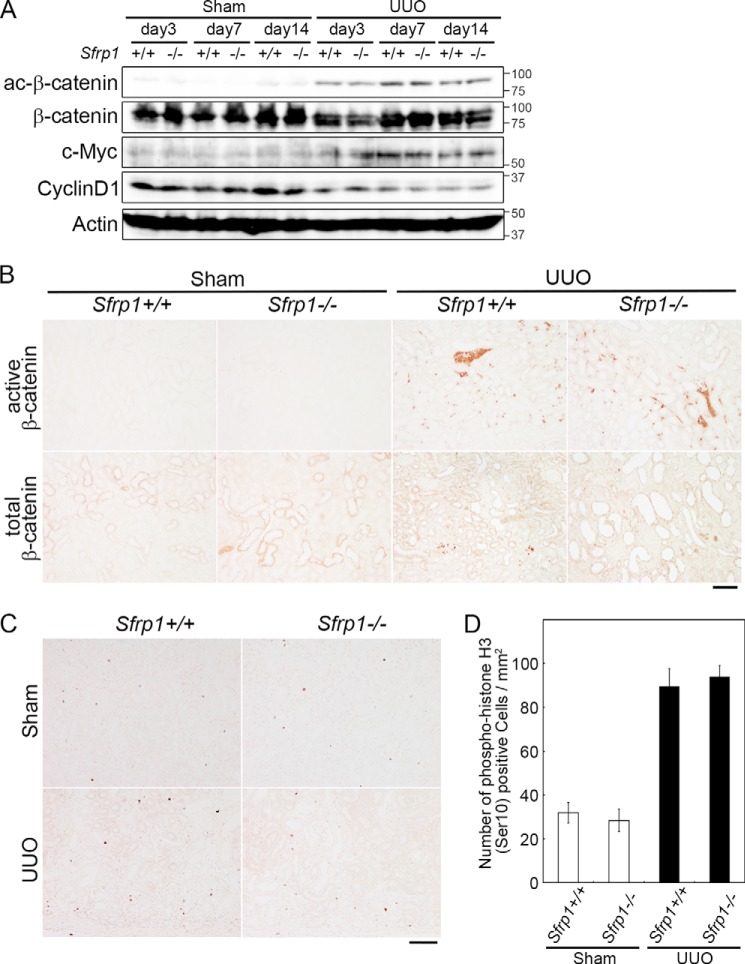
FIGURE 4.
Wnt/β-catenin pathway in the Sfrp deficient obstructed kidneys. A, detection of active- and total β-catenin, c-Myc, and cyclin D1 by Western blotting of the UUO kidneys. Actin was evaluated as an internal control. B, immunohistochemical analyses of mouse kidney sections with antibodies against active and total β-catenin in the sham and UUO kidneys at 7 days after the surgery. C and D, number of phospho-HistonH3 (Ser-10) cells in the obstructed kidneys at 7 days after UUO. C, section of the Sham and UUO kidneys in Sfrp+/+ and Sfrp−/− mice were subjected to immunostaining with an anti-phospho-histone H3 (Ser-10) antibody. D, graph shows analysis of the percentage of phospho-histone H3 (Ser-10)-positive cells in the UUO kidneys. Scale bars, 100 μm.

