FIGURE 6.
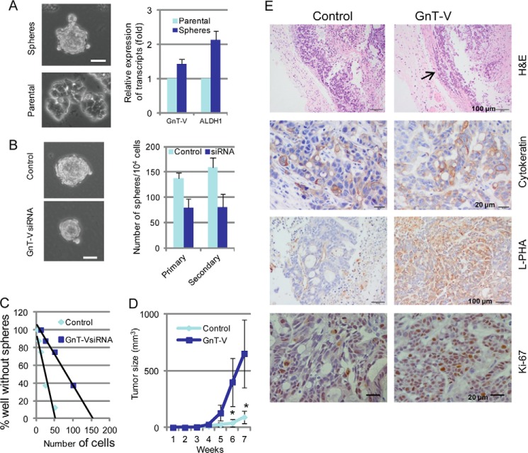
GnT-V regulates self-renewal and tumorigenicity of CCSC. A, HT-29 cells (4000 cells/well) were grown in stem cell culture media in suspension for 7–10 days, and tumorspheres were formed (left panel, top). Bar, 50 μm. Both aldehyde dehydrogenase-1 (ALDH1) and GnT-V mRNA levels were determined by qRT-PCR in both tumorspheres and parental cells on plastic plates (right panel). B, GnT-V-suppressed HT-29 cells (4000 cells/well) were grown in suspension for 7–10 days, and the number of tumorspheres was counted in 5–6 random fields. After tumorspheres were collected and single-cell suspension prepared, they (4000 cells/well) were grown in stem cell culture media in suspension for another 7–10 days for secondary sphere formation. Bar, 50 μm. C, primary tumorspheres formed from GnT-V suppressed HT-29 cells were dissociated and seeded in 96-well plates in stem cell culture media at densities ranging from 400 to 1 cell/well. After growth for 7–10 days, the percentage of wells that did not contain spheres at each cell plating density was calculated and plotted against the number of cells per well. D, Aldefluor-positive cells (3 × 10³) isolated from GnT-V overexpressing LS180 cells were injected subcutaneously into the backs of NOD/SCID mice (n = 5), and secondary tumor growth was observed for up to 8 weeks. *, p < 0.05. E, tumors were dissected at week 8, and H&E and other immunochemical staining were performed as indicated. The arrow indicates local invasion, whereas cytokeratin staining confirms the epithelial origin of the tumor cells.

