
Figure 5.
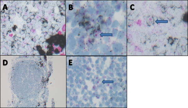
Non-specific nucleolar staining with lambda probe. A-C: Germinal center in a reactive tonsil. (A) polyclonal mixture of KAPPA and LAMBDA positive cells (400×); (B&C): arrows point out KAPPA expressing cells as evidenced by black ring of cytoplasmic staining with non-specific pink nucleolar staining (600×). D-E: Follicular lymphoma. (D) low power view of FL case which was indeterminate for KAPPA and lambda due to expressing neither light chain (40×); (E): high power view of same case demonstrating lack of cytoplasmic staining pattern for either KAPPA or LAMBDA light chain expression with non-specific pink nucleolar staining (600×).