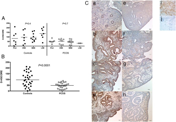
Figure 2.
A, Immunohistochemistry expression of phosphorylated GAB1 over the menstrual cycle in normal women (controls) and in women with PCOS. No significant difference was observed among different phases of menstrual either in controls (P = .4; ANOVA), or in women with PCOS (P = 0.7; ANOVA). B, Immunohistochemistry of endometrial expression of phosphorylated GAB1 between normal women (controls) and women with PCOS. A significant reduction was observed in endometrium of women with PCOS (P < .0001; unpaired Student t test). All phases of the menstrual cycle were grouped because no difference was found among phases within individual groups. Bars represent means. C, Representative images of immunostaining for phosphorylated GAB1 in endometrium of normal women (a, proliferative; b, early secretory; c, midsecretory; d, late secretory) and in women with PCOS (e, proliferative; f, early secretory; g, midsecretory; h, late secretory). Breast tissue was used as positive (i) and negative (j) external controls. Magnification is ×200. Bars represent 50 μm. ESE, early secretory endometrium; LSE, late secretory endometrium; MSE, midsecretory endometrium; Prol, proliferative phase.

