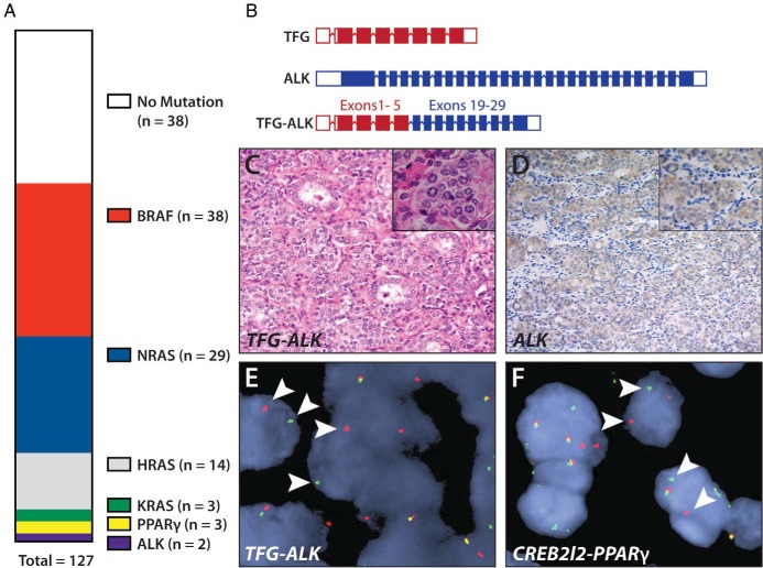
Figure 1.
Identification of oncogenic alterations in the FVPTC. A, Vertical parts-of-whole diagram of observed mutations in the panel of 127 FVPTC cases. B, Diagram of the TFG-ALK rearrangement observed in one tumor. C, Hematoxylin and eosin histology of the TFG-ALK rearranged tumor. D, Immunohistochemical detection of increased ALK expression in TFG-ALK rearranged tumor. E, FISH showing break-apart probes for ALK being separated in tumor cells (white arrowheads) in a TFG-ALK-positive tumor, indicating genomic translocation of ALK. F, FISH capture showing break-apart probes (white arrowheads) for PPARγ in a CREB3L2-PPARγ-positive tumor.

