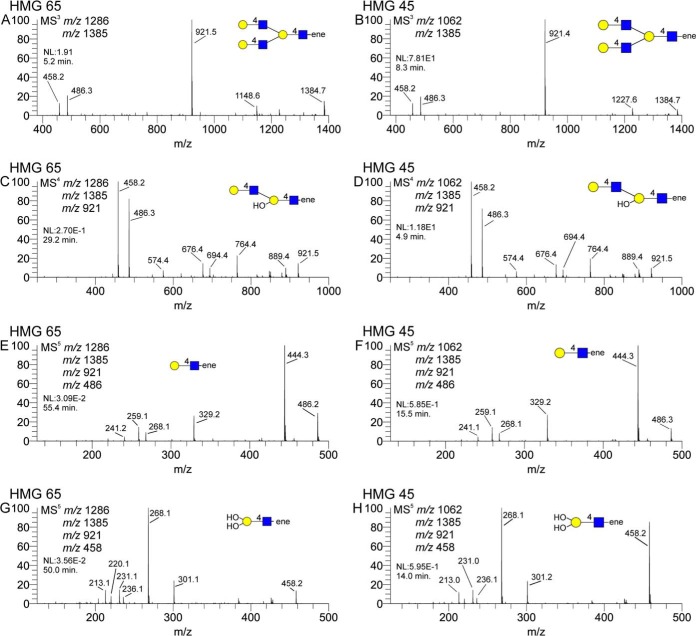
Fig. 9.
Isolation of a tri-LacNAc branching motif isolated from a previously described lacto-N-decaose and a previously undescribed octaose. Panels A and B show the MS3 spectra of the tri-LacNAc ion, panels C and D show the internal di-LacNAc component, panels E and F show the terminal LacNAc unit, and panels G and H show the branched internal LacNAc unit. Each spectral pair displays a remarkable degree of similarity for each HMG, suggesting a common substructure to each.

