Fig. 2.

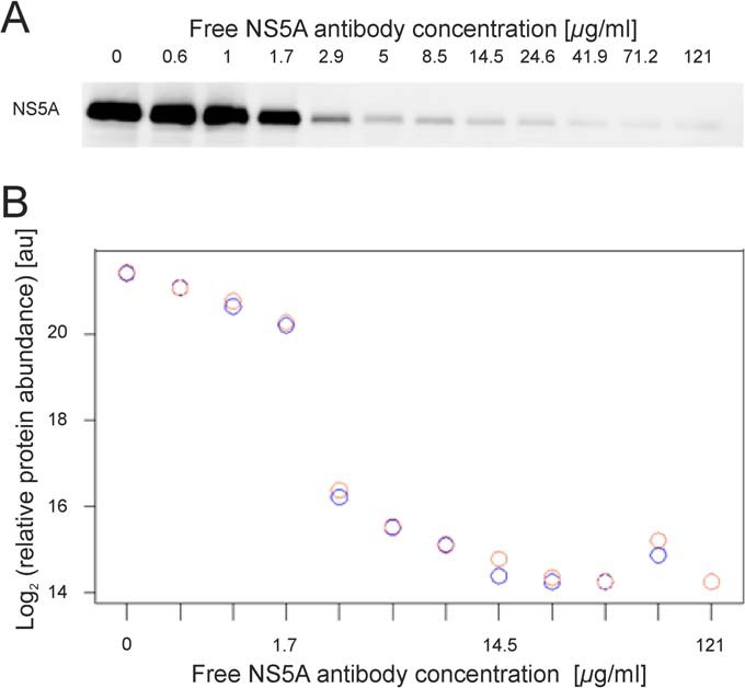
Concentration-dependent decrease of the NS5A capture by applying ICC-MS. A. NS5A capture from Con1-Huh7 cell lysates pre-incubated with 0, 0.6, 1, 1.7, 2.9, 5, 8.5, 14.5, 24.6, 41.9, 71.2, and 121 μg/ml of free NS5A antibody was probed by Western blot analysis. NS5A IP gradually decreased due to pre-incubation of the cell lysates with increasing concentration of the free form of the antibody. B. NS5A from each immunoprecipitate was identified and quantified by LC-MS. The relative abundance was plotted on the y axis and the free NS5A antibody concentrations on the x axis. Replicates are indicated by red and blue circles.
