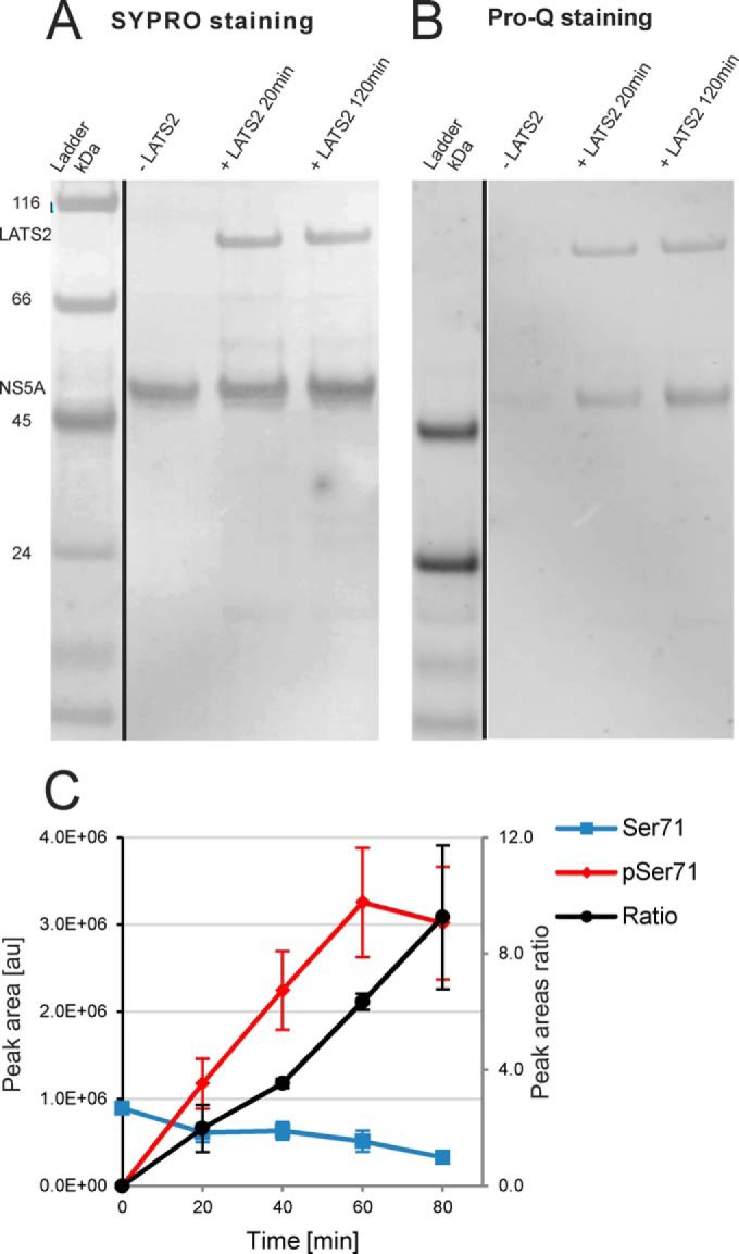
Fig. 4.

In vitro phosphorylation of NS5A by LATS2. A. Total protein (SYPRO) and B. phosphoprotein (Pro-Q Diamond) staining of the SDS-PAGE separated reaction mixtures. We used NS5A Δ32 (residues 33–447) recombinant form of the protein detected at ca. 50 kDa. We clearly observed phosphorylation of NS5A after 20 min treatment with the kinase. C. Relative quantitation of Ser71 phosphorylation by LC-MS. Ratios between phosphorylated and un-phosphorylated NGSMRIVGPK peptide EICs are represented. Error bars, S.E. (n = 2).
