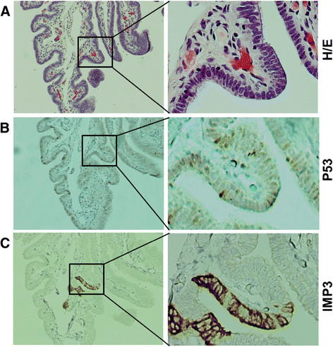
Figure 1.

Differential expression of IMP3 and p53 in normal tubal epithelial cells. A. H/E staining of normal epithelia of the fallopian tube. B. P53 was occasionally positive in some normal-looking secretory cells of the fallopian tube, which typically representing wild type TP53. C. IMP3 was strongly expressed in focal area of secretory cells in the fallopian tube, barely in ciliated cells in the only one case of the benign group. Ciliated cells could be appreciated by cilia on the left of panel A. Original magnifications: Left panel 40x, right panel 200x.
