Fig. 10.

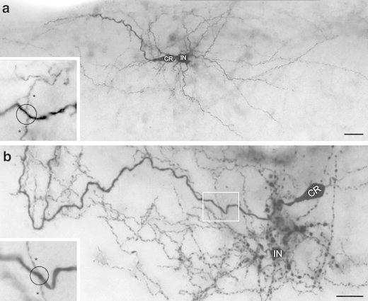
Light microscopy of CR cells and GABAergic interneurons in layer 1 of the neocortex. a, b Besides CR cells (CR), numerous GABAergic interneurons (IN) with different dendritic morphologies and axonal domains were found in layer 1. In both a and b one CR cell and one L1 interneuron is filled with biocytin. The interneuron in a displayed a multipolar elongated dendritic tree with a dense local axonal domain and individual long-range horizontal axonal collaterals confined to layer 1. The interneuron in b is of the neurogliaform type with local axonal collaterals and a high degree of collateralization and density of synaptic boutons. Insets in a, b High power light microscopy (framed area in b) suggests that the en passant axons (indicated by asterisks) of GABAergic interneurons establish synaptic contacts on proximal and distal dendrites (indicated by the open circle) of CR cells. Scale bars in a, b are 20 μm
