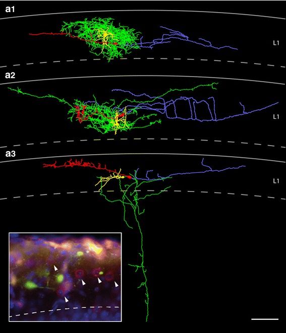
Fig. 11.

Neurolucida reconstructions of CR cells and GABAergic interneurons in layer 1 of the neocortex. a1–a3 Three representative examples of CR cells and adjacent GABAergic interneurons. The somatodendritic domain of the CR cells is given in red that of the interneurons in yellow, the axonal domains in blue (CR cells) and green (interneurons). The interneurons depicted in a and b display a local and dense axonal domain nearly completely covering the dendritic domain of the CR cells whereas the interneuron in c possess an axon projecting deep into layer 5 with collaterals in layer 2/3 and layer 4. Scale bar in a–c is 100 μm. Inset GABAergic interneurons in layer 1 as revealed by Gad67-immunoreactivity (indicated by arrowheads) are intermingled with EGFP-labeled CR cells
