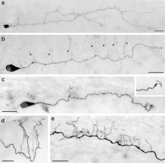
Fig. 2.

Dendritic morphology of neocortical CR cells. a–c Light microscopic images of three biocytin-filled CR cells from P11 old animals showing the heterogeneity in their dendritic morphology with respect to the length of the stem dendrite, side branches and frequency of spine-like, filopodial appendages. The CR cell in a shows a relatively high degree of collateralization and number of side branches covered with spine-like, filopodial appendages. The CR cell in b possessed a single thick stem dendrite with vertically oriented side branches (marked by asterisks); whereas the CR cell in c is an example with a short and smooth stem dendrite with only a single side branch. Inset: On dendritic side branches sometimes growth cone-like structures were observed. d High power photomicrograph of a vertically oriented tuft dendrite with side branches terminating near the pial surface. e Example of a CR cell stem dendrite with a high degree of short vertically, pial-oriented side branches. All figures are oriented such that the pial surface is on top. Scale bars in a–e are 20 μm
