Fig. 3.

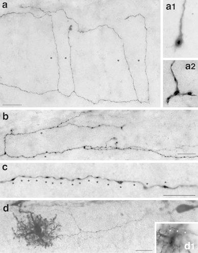
Axonal features of neocortical CR cells. a Axonal projection pattern of a typical CR cell with long vertically oriented axonal collaterals (marked by asterisks) spanning the entire volume between the pial surface and the layer 1/layer 2/3 border. Some of these collaterals still bear growth cones at their tips (a1, a2). b Axonal projection pattern of a typical CR cell with long horizontally oriented axonal collaterals, often running parallel to the main axon and the pial surface. c High power photomicrograph showing the high density and distribution of synaptic boutons (marked by asterisks) along a single axonal collateral. Note the different size of the synaptic boutons. d Dye- and synaptic coupling between a CR cell and a neighboring end foot astrocyte. Biocytin-filling of a single CR cell often led to co-labeling of astrocytes, but not of other L1 neurons or neurons in the underlying cortical layers 2/3 to 6. Note the invasion and ramification of the CR cell axon marked by asterisks into the astrocytic tree (d1). Scale bars in a, b, d are 20 μm and in c 10 μm
