Fig. 4.

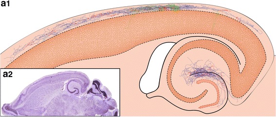
Location and distribution of all biocytin-filled and reconstructed CR cells and GABAergic interneurons in layer 1 of the neocortex. a1 Neurolucida reconstructions of all investigated and reconstructed CR cells and GABAergic interneurons in layer 1 of the neocortex. The somatodendritic domain of CR cells is given in red, their axonal arborization in blue. The somata and dendrites of the GABAergic interneurons are depicted in orange, their axons in green. CR cells formed a dense, horizontal network exclusively confined to layer 1 with individual CR cells spanning ~1.7 mm of cortical surface whereas most GABAergic interneurons form a more local plexus in layer 1 or project to the underlying cortical layers. In contrast, CR cells located in the dentate gyrus or str. lacunosum moleculare of the hippocampus always had two axonal domains, a dense local and a projection domain to various subregions of the hippocampus and even to the entorhinal cortex. a2 Nissl-stained section showing the plane of sectioning of the acute slices. For electrophysiological recordings slices were used in which the ‘barrel field’ of the somatosensory cortex and the hippocampal formation were visible
