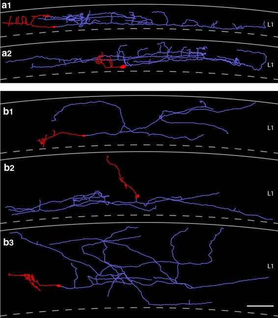
Fig. 5.

Neurolucida reconstructions of typical and ‘atypical’ CR cells in layer 1 of the neocortex. a1, a2 Two representative examples of CR cells showing the typical dendritic configuration and axonal arborization characteristic for CR cells. The somatodendritic domain is given in red, the axonal arborization in light blue. Top most lines indicate the pial surface, dashed lines the border between layer 1 and layer 2/3. b1–b3 Three representative examples of ‘atypical’ CR cells. These CR cells either showed alterations in their somatodendritic orientation and arborization (b2) or had a much broader axonal field spanning nearly the entire volume of layer 1 (b1, b3). Some color code as in a1 and a2. Scale bar in a1–b3 is 100 μm
