
Figure 1.
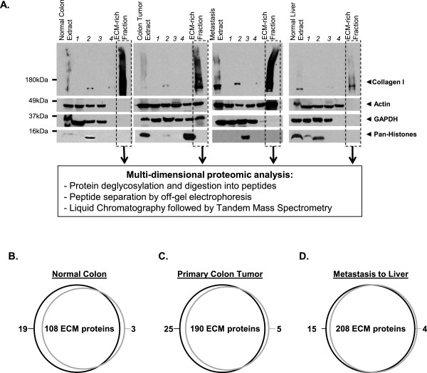
ECM protein enrichment from tissues and tumors and reproducibility of the proteomic analysis. A. The ECM protein enrichment and sequential extraction of intracellular components (steps 1 to 4) were monitored in each sample (normal colon, colon tumor, metastasis to liver and normal liver) by immunoblotting for collagen I (ECM marker), actin (cytoskeletal marker), GAPDH (cytosolic marker), and histones (nuclear marker). The insoluble fraction remaining after serial extraction was enriched for ECM proteins and largely depleted for intracellular components. B-D. Intra-patient reproducibility was assessed by comparing the ECM compositions of two distinct pieces of the same normal colon (B), primary colon tumor (C) or metastasis to liver (D) from patient 1. Venn diagrams show the intra-patient reproducibility in terms of matrisome proteins.