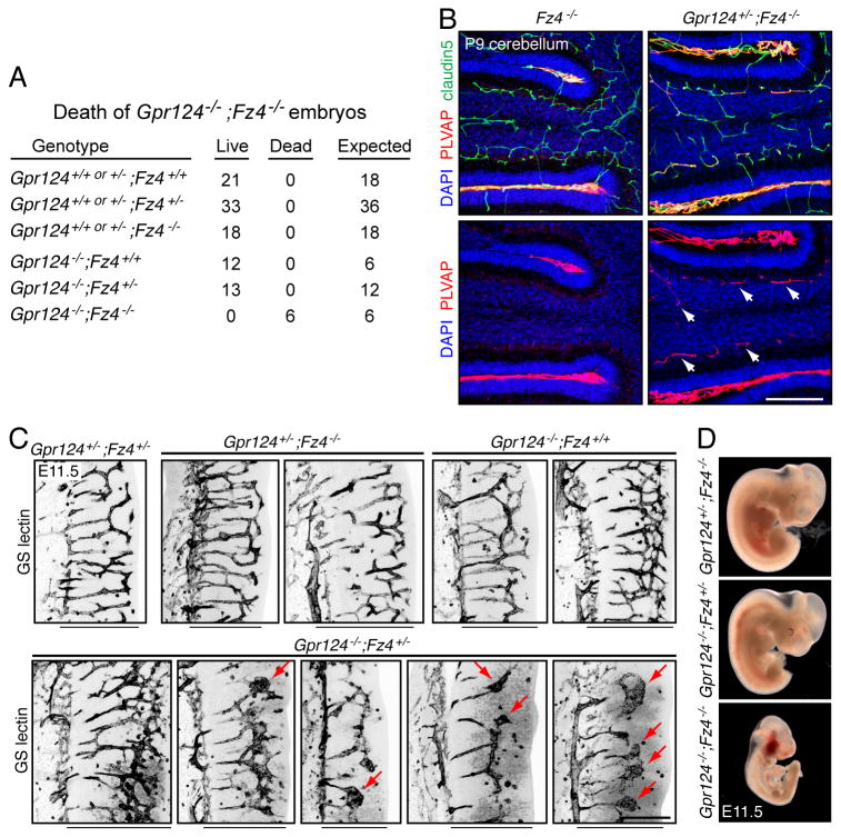
Figure 3. Gpr124 and Fz4 interact genetically in CNS angiogenesis and BBB maintenance.
(A) Genotypes and phenotypes of 97 embryos harvested at E11.5 from 15 timed pregnant females from a Gpr124+/-;Fz4+/- x Gpr124+/-;Fz4+/- intercross.
(B) Fz4-/- and Gpr124+/-;Fz-/- cerebella at P9. Gpr124+/-;Fz4-/- cerebella show accelerated conversion of granule cell layer ECs to a PLVAP+ state (arrows).
(C) Angiogenesis in E11.5 hindbrains. Within each image, the perineural vascular plexus is to the left and the ventricular surface is to the right. The width of the neuroepithelium is indicated by the black line beneath each image. Angiogenesis in Gpr124+/-;Fz4+/-, Gpr124+/-;Fz4-/-, and Gpr124-/-;Fz4+/+ hindbrains is indistinguishable from WT (upper panels). Angiogenesis in Gpr124-/-;Fz4+/- hindbrains is variably attenuated (lower panels). Hindbrain sections are shown from five Gpr124-/-;Fz4+/- embryos, and three show a severely impoverished vascular network with glomeruloid bodies (red arrows).
(D) Severe growth retardation of Gpr124-/-;Fz4-/- embryos at mid-gestation; all embryos were harvested at E11.5. Scale bars: 200 μm. See also table S2.

