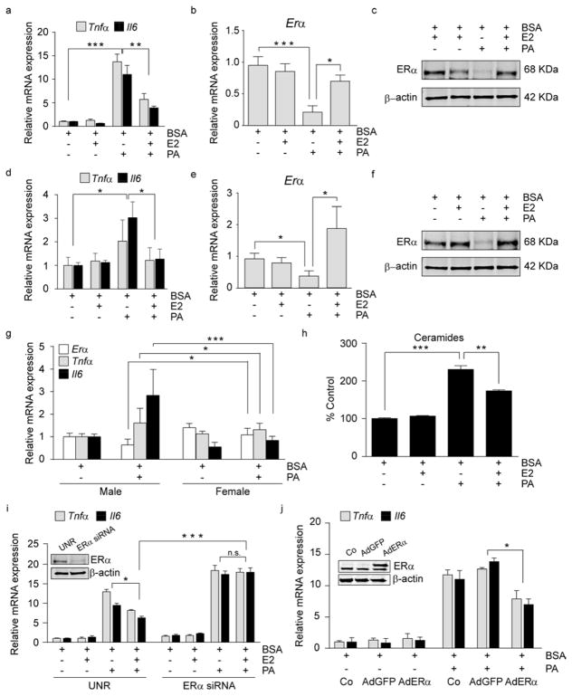
Figure 3. 17-β estradiol, through activation of ERα modulates PA-induced inflammation in hypothalamic neurons.
(a–c) N43 cells were pre-treated for 1 h with E2 and then treated for 8 h as indicated. (a) mRNA levels of Tnfα and Il6 in N43 cells. mRNA (b) and protein (c) levels of ERα in N43 cells following treatments. n=3. (d–f) Primary hypothalamic neurons were pre-treated for 1 h with E2 followed by 8 h of the indicated treatments. (d) mRNA levels of Tnfα and Il6 in primary hypothalamic neurons following the aforementioned treatments. mRNA of Erα (e) and ERα protein levels (f) in primary neurons. n=5. (g) mRNA levels of the indicated genes following 8 h treatment in primary male and female hypothalamic neurons. Males, n=5; females n=8. (h) Ceramides content in N43 cells following pre-treatment for 12 h with E2, where indicated, and treated for 8 h. n=3. (i) N43 cells were transfected with siRNAs, pretreated 48 h later for 1 h with E2, and cultured as indicated. mRNA levels of inflammatory markers following the treatments. The insert is a representative immunoblot of N43 cells transfected with control (UNR) siRNA or siRNA for ERα. n=3. (j) N43 cells were infected either with AdGFP (empty vector) or with AdGFP-ERα and then treated as indicated. Data represent mRNA levels of inflammatory markers following treatments. Insert shows representative immunoblots of N43 cells infected with the aforementioned viral constructs demonstrating ERα protein levels 48 h after the adenoviral exposure. n=3. Data are presented as mean ± SEM. *p< 0.05, **p< 0.01, ***p < 0.001. See also Figure S3.

