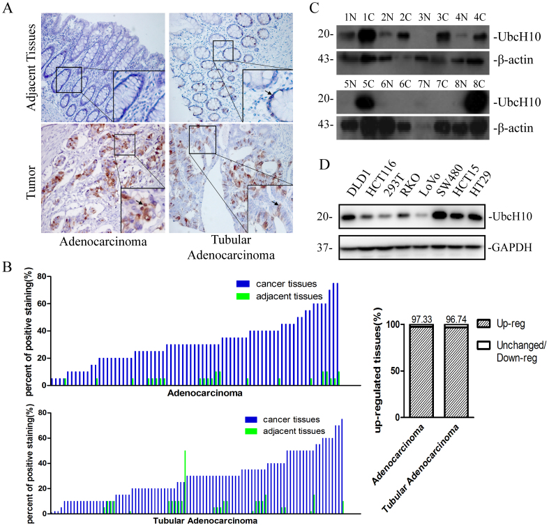
Figure 1. Expression of UbcH10 in colorectal cancer.
(A) Immunohistochemical staining of UbcH10 on tissue microarrays containing colorectal cancer tissues and adjacent normal tissues. Representative stainings of colon cancer tissues (bottom, left) and rectal cancer tissues (bottom, right) are shown. Arrows indicate the cells that stained positively. (B) Tissue microarray data analysis of UbcH10 expression in tumors and adjacent normal tissues from 75 patients with colon cancer and 92 patients with rectal cancer. Here shows the percent of positive staining cells in the corresponding tissue sample. (C) The UbcH10 protein expression levels of colorectal tumors and adjacent normal tissues were examined using Western blot. β-actin was used as a loading control. N represents normal tissue. C represents tumor tissue. (D) The protein expression levels of UbcH10 in 7 colorectal cancer cell lines and the 293T cell line were examined. GAPDH was used as a loading control.

