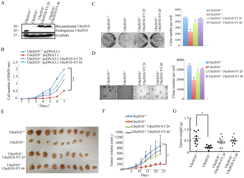
Figure 3. Rescue of repressed tumor formation via exogenous UbcH10 expression in vivo and in vitro.
(A) For the exogenous expression of wild-type recombinant UbcH10 in UbcH10-/- DLD1 cells, stable cell lines were generated via plasmid-mediated transfection. The expression of UbcH10 in the DLD1 cells was examined using Western blotting. GAPDH was used as a loading control. (B) Cell proliferation assay. Approximately 1 × 10³ cells of the indicated cell lines were seeded in 96-well plates and analyzed using the CCK8 method each day for seven consecutive days. Three repeats were performed and representative results are shown. (C) The indicated cells (3 × 10³) were seeded in 6-well plates and then cultured for approximately 10 days. The cell colonies were stained with crystal violet and photographed. The clones were counted and plotted. (D) Soft agar colony formation assay. The indicated cells (1 × 10⁴) were seeded in 6-well plates with 0.35% upper agar and 0.7% lower agar for 14 days. The cell colonies were photographed, counted, and plotted. (E–G) Xenograft experiments were performed by injecting the indicated cells (5 × 10⁶) into the flanks of 6-week-old nude mice to form tumors. The tumors were measured every 3 days, and the tumor volume was calculated using the formula ([length × width2] × 0.5). The tumors were removed and weighed 23 days after injection. The numbers of colonies are expressed as the means ± S.E. from six assays. *P< 0.05, **P< 0.01, ***P< 0.001 compared with controls.

