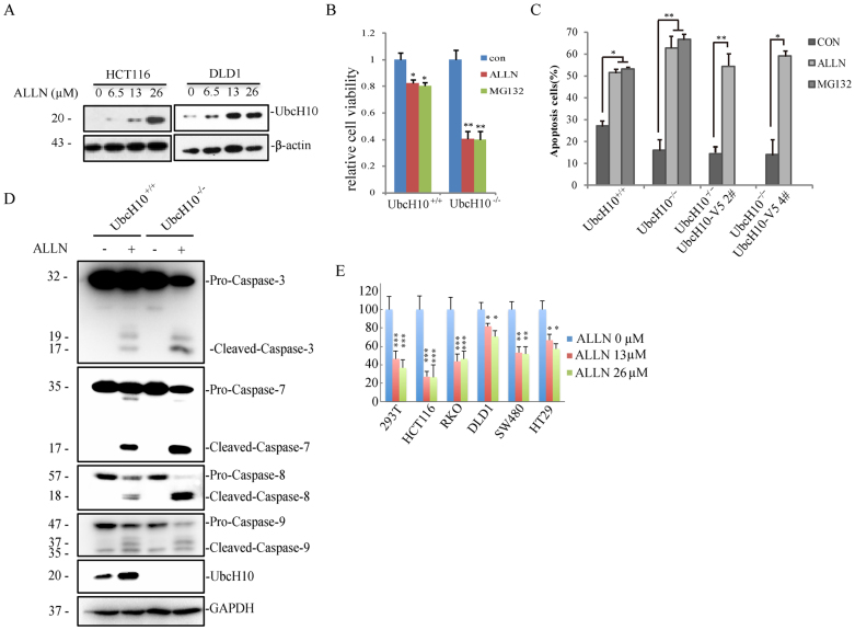
Figure 6. UbcH10 deficiency aggravates ALLN-induced apoptosis.
(A) HCT116 and DLD1 cells were exposed to the indicated concentrations of ALLN for 24 hours. The cell lysates were analyzed via immunoblotting using anti-UbcH10 antibody. β-actin was used as a loading control. (B) DLD1-UbcH10–/– cells are sensitive to both ALLN- and MG132-induced apoptosis. Approximately 1 × 10⁴ cells of the indicated cell lines were seeded in 96-well plates and treated with 26 μM ALLN or 3.3 μM MG132 for 24 hours. The viability of the cells was measured immediately using the CCK8 method. (C) Annexin-V/PI staining followed by flow cytometry. DLD1 and DLD1-UbcH10–/– cells were exposed to ALLN (26 μM) or MG132(3.3 μM) for 24 hours. The cells were then stained with Annexin-V/PI for 15 min, and apoptosis was analyzed via flow cytometry. The early and late apoptosis rates of the cells are shown. (D) DLD1 and DLD1-UbcH10–/– cells were treated with ALLN (0 or 26 μM) for 24 hours. The cell lysates were analyzed via immunoblot using the indicated antibodies. GAPDH was used as a loading control. (E) Cell susceptibility to ALLN is related to UbcH10 level. Approximately 1 × 10⁴ cells of the indicated cell lines were seeded in 96-well plates and treated with various concentrations of ALLN for 24 hours. The viability of the cells was measured immediately using the CCK8 method. The cell numbers are expressed as the means ± S.E. from six assays. *P< 0.05, **P< 0.01, ***P< 0.001 compared with controls.

