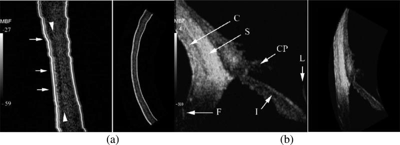
Fig. 20.
(a) LASIK–treated cornea six years post–treatment imaged at 75 MHz. Small arrows indicate various discontinuities in Bowman's layer, at the interface of the 50 μm thick epithelium and the underlying stroma. The stroma anterior to the ablation interface is approximately 2–3 dB lower in mean backscatter than the posterior residual stroma. (b)

