
Figure 5.
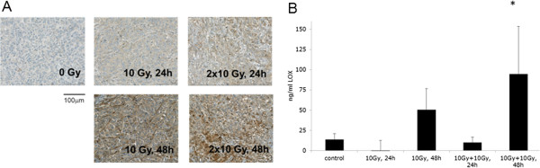
IR-enhanced LOX in tumor xenografts. (A) Tumor tissue from A549-derived tumor xenografts treated with 1 × 10 Gy and 2 × 10 Gy (with 12 h between fractions) and evaluated by immunohistochemistry for LOX at different time points after irradiation. Whole tumor sections were quantified for specific LOX-staining intensity. Each treatment group consists of 3 animals. At least 3 sections per tumor/animal were analyzed. (B) LOX in murine blood serum derived from mice with untreated or locally irradiated A549 tumor xenografts and quantified by ELISA at different time points after irradiation. Each treatment group consists of 3 animals.