Figure 3.

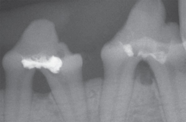
Periapical radiograph of the left mandibular second and third premolars sealed with gutta-percha and AH Plus, respectively, showing presence of periodontal lesion in the perforation region

Periapical radiograph of the left mandibular second and third premolars sealed with gutta-percha and AH Plus, respectively, showing presence of periodontal lesion in the perforation region