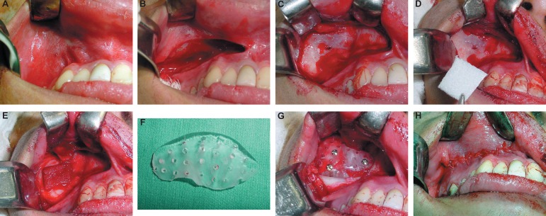
Figure 4.
Panel of intraoperative photographs. A: Access to the zygomatic area. B-C: Incision in the superior left labial vestibule. D-E: Fibrin sponge placed in this area. F: Prosthesis recontoured by trimming it with an acrylic bur. G: Prosthesis fixed with three titanium screws. H: Mucoperiosteal flaps repositioned and closed with Vicryl sutures.

