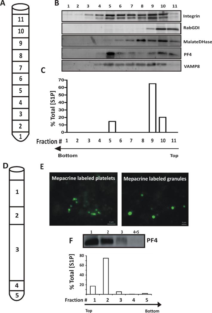
Figure 5. Localization of S1P to platelet granules.
(A) Human platelets were disrupted by nitrogen cavitation and the material was layered onto a continuous, sucrose gradient (30-60% (w/v)) and organelles were separated by ultracentrifugation (n=4). 1 mL fractions were collected as indicated. (B) TCA precipitated proteins from each fraction were probed by western blotting for the indicated markers. (C) Lipids were extracted from the corresponding fractions and S1P was measured by HPLC-MS/MS. S1P in each fraction is expressed as percent of Total S1P in the starting platelets. The data are representative of three independent experiments. (D) Platelets were incubated with mepacrine to label dense granules before disruption by sonication and fractionation of the material obtained by layering onto a 35%, 38% metrizamide step gradient and ultracentrifugation. The fractions were harvested as indicated: Fraction 1, 500 μL; 2, 200 μL; 3, 1.2 mL; 4, 100 μL; 5 (pellet), 100 μL (n=4). (E) Mepacrine-labeled, whole platelets and Fraction 5, containing platelet dense granules were imaged by epifluorescence microscopy. (F) Lipids were extracted from the gradient fractions and their S1P content was measured by HPLC-MS. The values are represented as percent of Total [S1P] in each fraction. The corresponding fractions were TCA precipitated to detect PF4 by western blotting. The data are representative of three different experiments.

