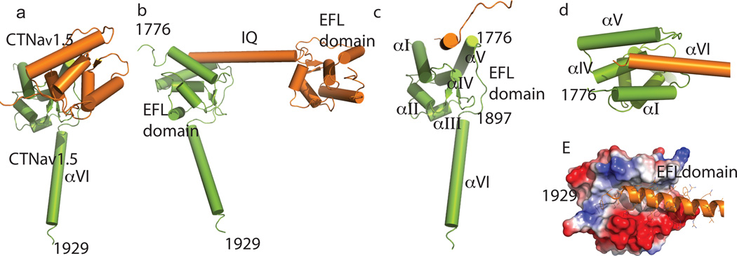
Figure 3. Interaction of the CTNav1.5 EFL domain with a neighboring CTNav1.5 helix αVI.
(a) Front view of the CTNav1.5-CTNav1.5 dimer. Residues of one CTNav1.5 molecule (green) interact with another CTNav1.5 molecule (orange). (b) Same as panel A but rotated 90 ° from A in the plane of the figure. (c)90 ° rotation from panel B showing the concave cavity formed by the helices of EFL domain; helix αI 1788–1801; helix αII 1814–1820; helix αIII 1832–1837; helix αIV 1850–1858; helix αV 1866–1882; helix αVI 1897–1926. (d) Close up of the cavity formed by helices αI and αV with helix αVI. (e) Same as D but with one CTNav1.5 colored according to the electrostatic potential. It displays a hydrophobic surface with ionic patches at the beginning and end.

