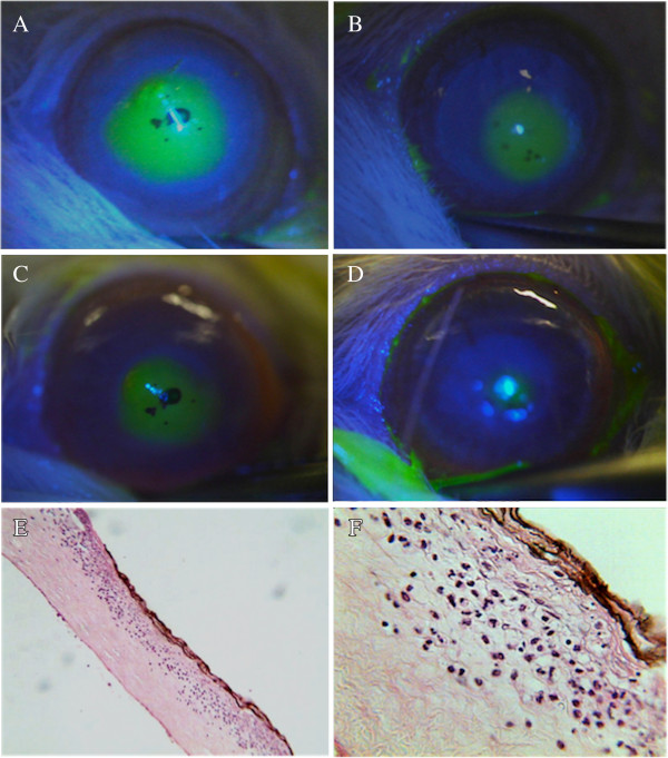
Figure 1.

Photograph composition of representative images of corneas from rabbits of the Brazilian propolis (BP) and vehicle (VH) groups. A to D - comparison of the fluorescent-stained area of injured cornea between groups at different time points, as following: A and B - photographs at 12 hours of animals from VH and BP groups respectively; C and D – photographs at 48 hours of animals from VH an BP groups respectively. Note the smaller areas of injury with BP treatment (images B and D). Image E displays a representative histological section of central cornea of BP group at 24 hours (H&E; original magnification 100X), and F shows details of this area, including a brownish central lesion with loss of epithelium and superficial stroma, necrosis and infiltration of neutrophils (H&E; original magnification: 400X).
