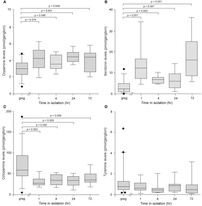
Figure 2.
Analysis of the levels of four biogenic amines isolated from individual metathoracic ganglia using HPLC with electrochemical detection. (A) Box and whisker plots showing median, interquartile range, and minimum and maximum values of the levels of dopamine in the metathoracic ganglion that increased significantly in locusts after only 1 h in isolation compared to gregarious controls. (B) Serotonin levels increased significantly in the metathoracic ganglion after 1 h in isolation. (C) By contrast the levels of octopamine decreased significantly after 1 h in isolation. (D) The levels of tyramine showed no difference between gregarious and isolated locusts. Results are based on n = 9–11 animals tested at each time point for isolated and long-term gregarious groups and significance tested using Kruskal–Wallis, followed by pair-wise Mann–Whitney U-tests, indicated above each graph.

