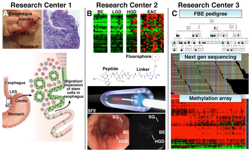
Fig. 2. Scientific focus of Research Centers.
A) Research Center 1 is studying the possible origins of BE and EAC using the pL2-IL-1β mouse model which develops multiple tumors at the squamocolumnar junction (SCJ) at 12 months. Photo and histology of pL2-IL-1β mouse with esophagitis and a tumor at the SJC at 15 months are shown. Schematic illustration of migrating stem cells (green, Lgr5+) at the cardia that expand into the inflamed esophagus. B) Research Center 2 is developing a molecular imaging approach by identifying targets that are gene amplified and/or highly overexpressed. Highly specific peptides are selected and fluorescently-labeled for in vivo imaging with a multi-spectral scanning fiber endoscope that can detect multiple targets simultaneously. This “red-flag” approach will help physicians to identify HGD in BE. C) Research Center 3 is investigating susceptibility genes in Familial Barrett’s Esophagus (FBE) and the role of methylated genes in BE and EAC. Pedigree shows affected subjects with BE (blue) and EAC (red). Alignment of reads from next generation sequencing run. Heat map from methylation arrays and non-coding and coding RNA signatures in BE and EAC.

