Figure 2.
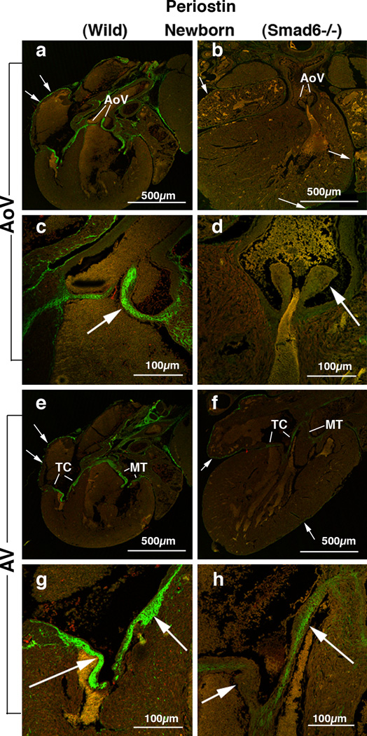
Periostin localization in Smad6+/+ (wild type) (panels a, c, e and g) and Smad6−/− littermate (panels b, d, f and h) newborn mouse aortic (AoV) and AV (AV) valves.
(a) Intense periostin expression is shown in aortic valves (AoV) in a Smad6+/+ (wild type) newborn mouse heart. Arrows indicate periostin expression in the epicardium surrounding the atrial wall.
(b) Periostin expression in a Smad6−/− newborn mouse heart. Periostin is localized in the epicardium (arrows); however, only weak immunostaining is observed in the aortic valves (AoV).
(c) Higher magnification view of the aortic valves (arrow) in panel a, showing intense expression of periostin.
(d) Higher magnification view of the aortic valves (arrow) in panel b. Periostin protein expression in the aortic valves is diminished in a Smad6−/− newborn mouse heart.
(e) Intense periostin expression is shown in the tricuspid (TC) and mitral (MT) valves in a Smad6+/+ (wild type) newborn mouse heart. Arrows indicate periostin expression in the epicardium.
(f) Periostin expression in a Smad6−/− newborn mouse heart. Periostin is localized in the epicardium (arrows); however, weaker immunostaining is observed in the tricuspid (TC) and mitral (MT) valves in a Smad6−/− newborn mouse heart than in the wild type heart.
(g) Higher magnification view of the tricuspid valves (arrows) in panel e, showing intense expression of periostin.
(h) Higher magnification view of the tricuspid valves (arrows) in panel f, showing weaker periostin immunostaining in the Smad6−/− heart than in the wild type heart.

