Figure 4.
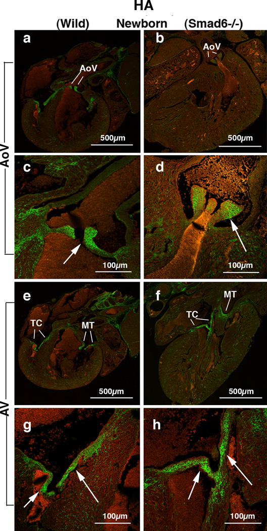
Hyaluronan (HA) deposition in Smad6+/+ (wild type) (panels a, c, e and g) and Smad6−/− littermate (panels b, d, f and h) newborn mouse aortic (AoV) and AV (AV) valves. HA localization is detected by using hyaluronan-binding protein (HABP).
(a) Intense HA deposition is shown in the aortic (Ao) valves in a Smad6+/+ (wild type) newborn mouse heart.
(b) HA localization in a Smad6−/− newborn mouse heart. HA immunostaining is evident but weaker in the Smad6−/− aortic valves (Ao) compared to that in the wild type aortic valves.
(c) Higher magnification view of the aortic valves (arrow) in panel a, showing intense expression of HA.
(d) Higher magnification view of aortic valves (arrow) in panel b. HA expression in the aortic valves is slightly reduced in a Smad6−/− newborn mouse heart.
(e) Intense HA deposition is shown in tricuspid (TC) and mitral (MT) valves in a Smad6+/+ (wild type) newborn mouse heart.
(f) HA localization in a Smad6−/− newborn mouse heart. HA is localized in tricuspid (TC) and mitral (MT) valves. (g) Higher magnification view of the tricuspid valves (arrows) in panel e, showing intense expression of HA in tricuspid valves (arrows).
(h) Higher magnification view of the tricuspid valves (arrows) in panel f. HA deposition in a Smad6−/− tricuspid valves appears similar to that of wild type newborn mouse.

