Figure 3.
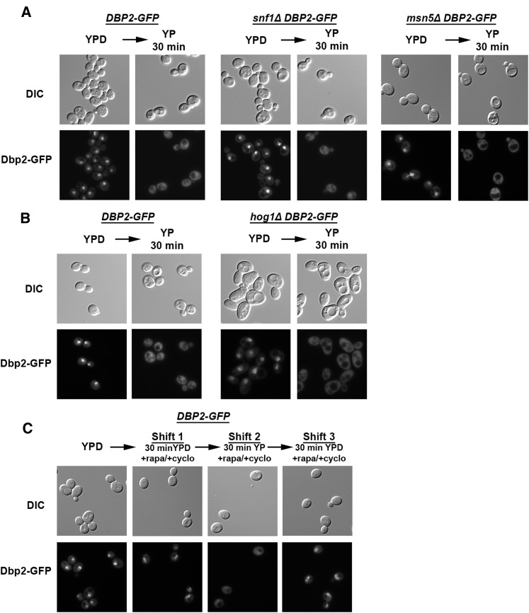
The cellular redistribution of Dbp2 upon glucose deprivation does not depend on the Snf1 pathway, Hog1 pathway, or TOR signaling. (A) Dbp2–GFP signal relocalization is not dependent on the Snf1 kinase pathway. Wild-type, snf1∆, and msn5∆ cells harboring genomically encoded DBP2–GFP constructs were visualized for Dbp2 localization in the presence of glucose (YPD) or after a 30-min deprivation (YP 30 min). Dbp2–GFP was visualized by epifluorescent microscopy and images are representative of three biological replicates. (B) Dbp2–GFP signal is not redistributed to the cytoplasm via the HOG1 osmolaric stress response pathway. Dbp2–GFP localization in wild-type and hog1∆ cells was visualized in both the presence of glucose (YPD) and following a 30-min glucose deprivation (YP 30 min) as above. (C) Glucose-dependent localization of Dbp2–GFP is not dependent on the TOR pathway. Dbp2–GFP cells were grown in the presence of glucose (YPD) and then shifted to YPD supplemented with rapamycin for 30 min to inhibit the TOR pathway and cycloheximide to inhibit de novo protein synthesis (Shift 1). Cells were then subjected to a 30-min glucose deprivation (YP 30 min) in the presence of rapamycin and cycloheximide (Shift 2). After glucose deprivation, cells were given access to glucose (YPD) supplemented with rapamycin and cycloheximide (Shift 3). Fluorescent images were collected as above.

