Figure 1.
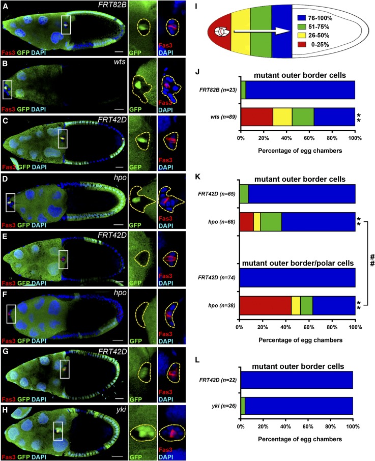
hpo and wts are required for border cell migration. GFP-negative mitotic clones were generated in FRT82B (A), FRT82B wtsX1 (B), FRT42D (C and E), and FRT42D hpo42-47 (D and F) and examined 6 days after clone induction. Mitotic clones of FRT42D (G) and FRT42D ykiB5 (H) were examined 3 days after clone induction. The ovaries were immunostained with anti-Fas3 and anti-GFP antibodies. Cell nuclei were stained with DAPI. Stage-10 egg chambers were selected and oriented as anterior toward the left. The border cell cluster is composed of two Fas3-positive polar cells in the center surrounded by four to six outer border cells. High magnification views of border cell clusters are shown in the panels on the right in A–H. (A) A border cell cluster containing GFP-positive polar cells and GFP-negative FRT82B control outer border cells migrated normally and reached the oocyte-nurse-cell border. (B) A border cell cluster containing GFP-positive polar cells and GFP-negative wts mutant outer border cells failed to migrate. (C) A border cell cluster containing GFP-positive polar cells and GFP-negative FRT42D control outer border cells reached the oocyte-nurse-cell border. (D) A border cell cluster containing GFP-positive polar cells and GFP-negative hpo mutant outer border cells failed to migrate. (E) A border cell cluster containing GFP-negative FRT42D control polar cells and outer border cells reached the oocyte-nurse-cell border. (F) A border cell cluster containing GFP-negative hpo mutant polar cells and border cells failed to migrate. (G) A border cell cluster containing GFP-positive polar cells and GFP-negative FRT42D control outer border cells reached the oocyte-nurse-cell border. (H) A border cell cluster containing GFP-positive polar cells and some GFP-negative yki mutant outer border cells migrated >75%. (I) A diagram demonstrates colors representing the distance of border cell migration. (J) Quantification and percentage distribution of border cell migration. Only border cell clusters with GFP-positive polar cells and GFP-negative outer border cells were counted. wts mutation in outer border cells severely impaired migration. (K) Border cell clusters were categorized into two groups. The group with mutant outer border cells contained GFP-positive polar cells and GFP-negative mutant outer border cells; the group with mutant outer border/polar cells contained one or two GFP-negative mutant polar cells and GFP-negative mutant outer border cells. The migratory defect was more severe in the group with mutant outer border/polar cells than it was in the group with mutant outer border cells. (L) Only border cell clusters with GFP-positive polar cells and some GFP-negative outer polar cells were counted. yki mutation did not affect migration 3 days after clone induction. Wilcoxon rank-sum test, **P < 0.01 comparing with control; ##P < 0.01. Bar, 20 μm in A–H.

