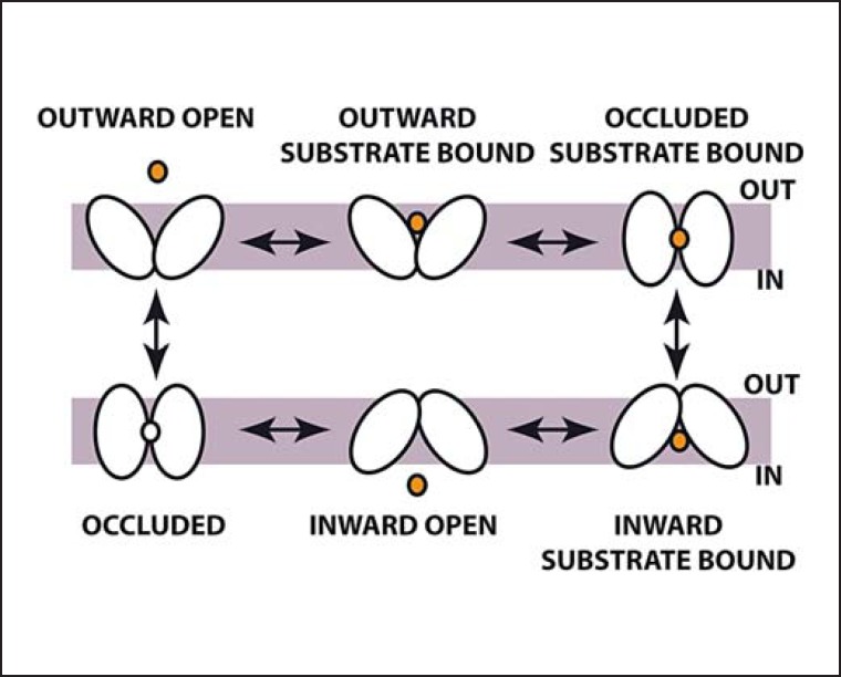
Fig. 2.
Topology diagrams along with experimental structures of transmembrane transporters. The ASBT family has ten transmembrane helices (TMH). Substrate (star) is bound between two interrupted helices, TMH4 and TMH9. MFS proteins are arranged as two bundles of six consecutive TMH each, which form a central substrate-binding site at the interface of the two bundles (amino acids in MCT8 participating in transport are indicated). LeuT fold transporters are arranged as two 5-helix inverted repeats followed by two TMH which do not participate in pseudo-symmetry. The substrate-binding site is formed by two interrupted helices, TMH1 and TMH6. The helices are colored from violet (helix 1) to red (helix 12).

