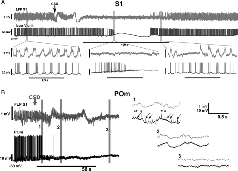
Figure 4.
Response of intracellular L5 and POm activity to the altered cortical activity. (A) Cortical LFP (top row, light gray) and intracellular activity of a rat L5 cortical neuron (bottom row, dark gray) is shown before, during and after a cortical spreading depression (arrow at CSD onset) induced by 2 M KCl in the primary somatosensory cortex (BC). Periods indicated by gray bars are shown below on an extended time scale. Before the CSD (left) L5 neurons display rhythmic spiking activity and depolarizing membrane potential oscillation locked to the Up states of the LFP. During the CSD (middle), the rhythmic L5 activity is first replaced by a ramp depolarization and high-frequency tonic firing (depolarizing phase) then by a depolarization blockade and a complete cessation of firing. After the CSD (right), L5 activity resumes. The time difference between the onset of CSD in the LFP and in the intracellular activity is the result of the propagation of spreading depression from the site of induction (LFP recording electrode) to the site of intracellular recording. (B) Cortical LFP (top row, gray) and intracellular activity of a POm neuron (bottom row, black) before and during CSD induced in the primary somatosensory cortex (S1). Periods indicated by gray bars (1–3) are shown on an extended time scale on the right. During the depolarization phase of the CSD, when L5 neurons display high-frequency tonic firing (see A) the POm neuron receives a barrage of high-frequency EPSPs (1). In the next phase of the CSD (2–3), when L5 neurons stop firing, all EPSPs disappear in POm.

