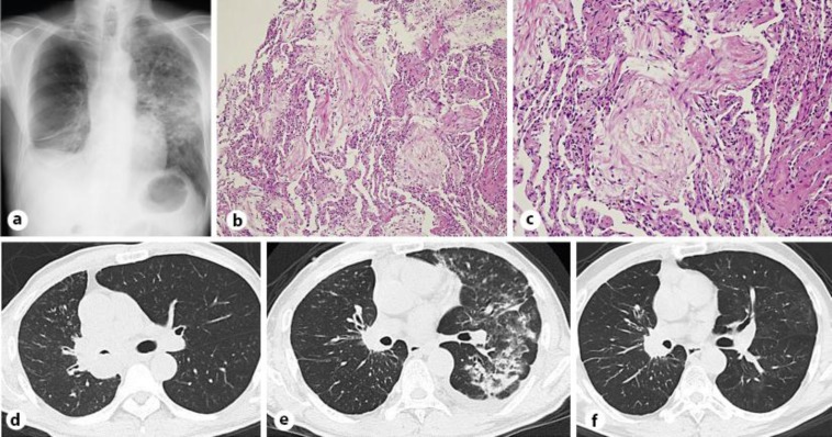
Fig. 1.
a Chest radiograph showed a unilateral infiltrate in the middle lung field of the left lung. b Transbronchial lung biopsy specimen from the left lower lobe showed intra-alveolar granulation tissue with myofibroblasts consistent with OP. HE. ×100. c Mass on body was seen on the transbronchial lung biopsy specimen. HE. ×200. d Chest CT scan showed a tumor lesion in the right pulmonary hilum and the narrowed right main bronchus. e Chest CT scan revealed panlobular lesions in the left lung. f Repeat chest CT scan showed a significant improvement of parenchymal changes 8 weeks after starting corticosteroid therapy without recurrence of OP.

