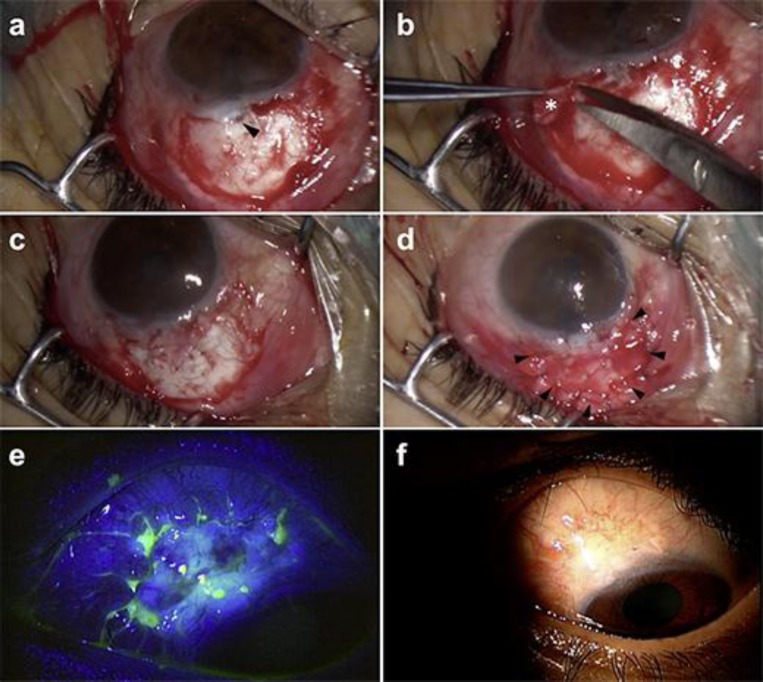
Fig. 2.
a After the avascular bleb tissue and the melted scleral flap had been removed, the scleral fistula (arrowhead) can be seen. b Thick fibrotic tissue is carefully separated in a sheet of membrane (*) that is used for the autologous Tenon's graft. c The graft is cut to the desired size to cover the defect and sutured with 10-0 nylon sutures. d A layer of amniotic membrane (arrowheads) is applied over the exposed sclera. e Two weeks postoperatively, the site is totally re-epithelialized with no aqueous leak. f Three months postoperatively, vascularization into the surgical site is observed.

