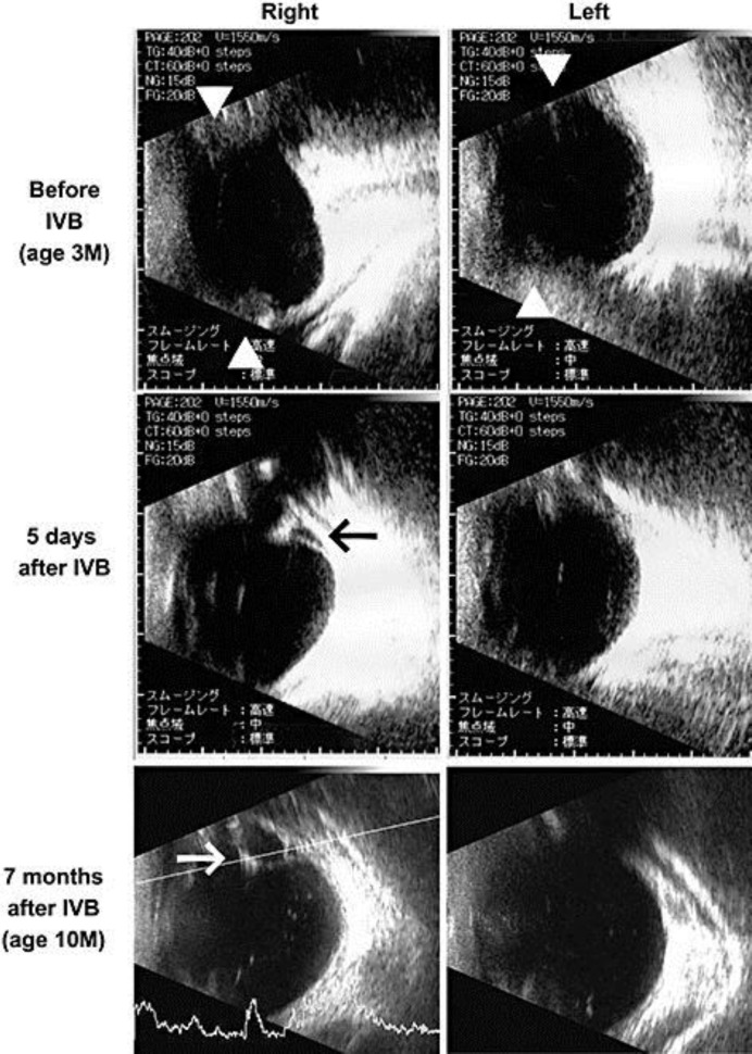
Fig. 2.
Ultrasonographic findings of eyes with Peters anomaly and ROP. Before the IVB, abnormal echo was detected which suggested ridge formation (triangles in the upper row, stage 3 ROP). Five days after the IVB, a retinal detachment was suspected in the right eye (black arrow in the middle row, stage 4A ROP). Seven months after the IVB, a third retinal detachment is suspected in the right eye (white arrow in the lower row, stage 4A ROP).

