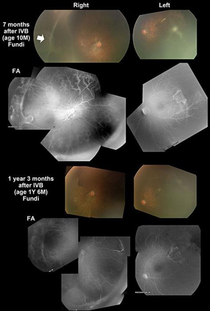
Fig. 3.
Fundus photographs and fluorescein angiograms. A retinal detachment can be seen in the right eye 7 months after the IVB (arrow). An avascular zone and some leakage of fluorescein are still present in the periphery 1 year and 3 months after IVB. Photography was difficult because of the residual corneal opacity in both eyes. Fundus photography and fluorescein angiograms were performed using RetCam® 3.

