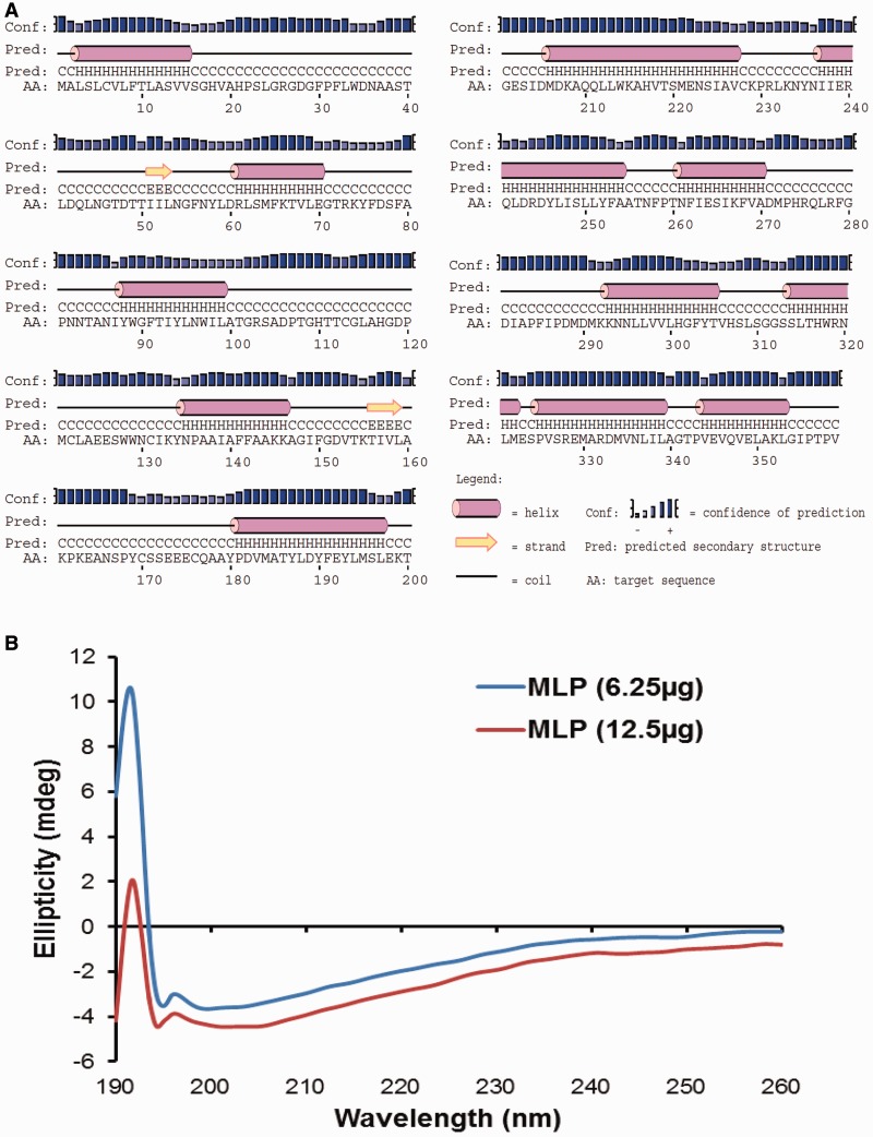
Fig. 8.—
MLP secondary structure analysis. MLP is an α-helical protein determined by PSIPRED prediction followed by CD signal in the far-UV region. (A) Platypus MLP secondary structure predicted by PSIPRED server shows high proportion of alpha helix content. Helical and β-sheet secondary structure elements are depicted by pink barrels and yellow arrows, respectively. Prediction confidence is represented by blue bars. (B) Platypus MLP secondary structure analysis determined by CD. The CD spectra acquired in the range of 190–260 nm for recombinant MLP. Analysis of CD spectrum shows a positive dichroic band with a maximum at 190 nm that highlights the content of α-helices in the protein.

