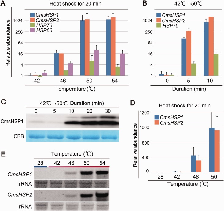
Fig. 4.—
Expression patterns of CmsHSP1 and CmsHSP2. (A) Expression profiles of CmsHSP1, CmsHSP2, HSP60, and HSP70 in cells cultured at 42 °C. The cDNAs were prepared from cells cultured at 42 °C and exposed to a heat shock at 46, 50, or 54 °C for 20 min. Relative accumulation levels were normalized to the levels for the cells at 42 °C. (B) Changes in the mRNA levels of CmsHSP1, CmsHSP2, and HSP70 during heat shock treatment obtained by qRT–PCR. The cDNAs were prepared from cells cultured at 42 °C and exposed to heat shock at 50 °C for 5 and 10 min. (C) Western blot showing the accumulation of CmsHSP1 protein from 5 to 30 min. Coomassie Brilliant Blue is a loading control. (D) Expression profiles of CmsHSP1 and CmsHSP2 in cells cultured at 28 °C. The cDNAs were prepared from cells cultured at 28 °C and exposed to heat shock at 42, 46, or 50 °C for 20 min. Relative accumulation levels were normalized to the levels for the cells at 28 °C. (E) Northern blot showing the expression profiles of CmsHSP1 and CmsHSP2. rRNA is a loading control. Total RNA was prepared from cells cultured at 28 °C and exposed to heat shock at 42, 46, 50, or 54 °C for 20 min.

