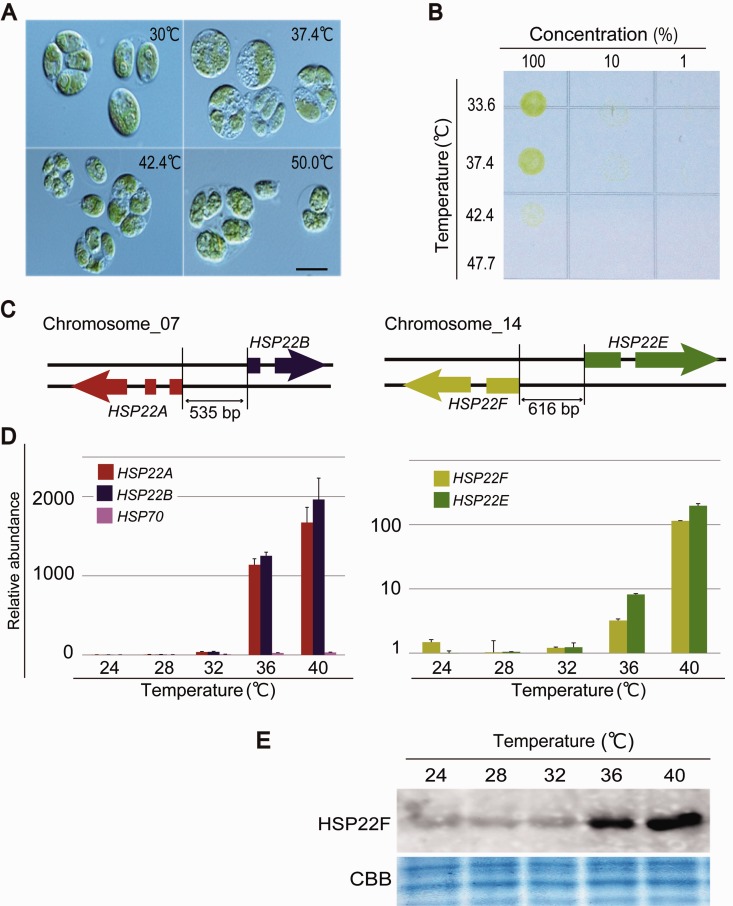
Fig. 5.—
Effect of heat shock on the green alga Ch. reinhardtii. (A) Microscopic images of the cells exposed to a heat shock for 20 min. Bar is 5 µm. (B) Survival rate of cells exposed to a heat shock for 20 min. Numbers across the top indicate the relative quantity of cells originally plated on TAP medium. (C) Schematic representation of HSP22F and HSP22F located in tandem on the chromosome. (D) Expression profiles of HSP22A, HSP22B, and HSP70, and of HSP22F and HSP22E. (E) Western blot showing the accumulation of the HSP22F protein in cells exposed to a heat shock for 20 min.

