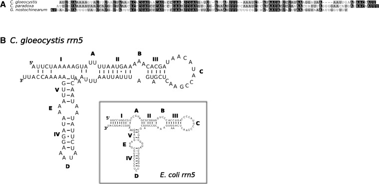
Fig. 2.—
Putative rrn5 sequence from Cyanoptyche gloeocystis mtDNA, aligned with rrn5 sequences from C. paradoxa and G. nostochinearum (A); putative secondary structure of Cyanopt. gloeocystis rrn5 (B). The inset shows the proposed structure of the E. coli rrn5 for reference. Helices are shown by Roman numerals (I–V), and single-stranded regions are shown by upper case letters (A–D).

