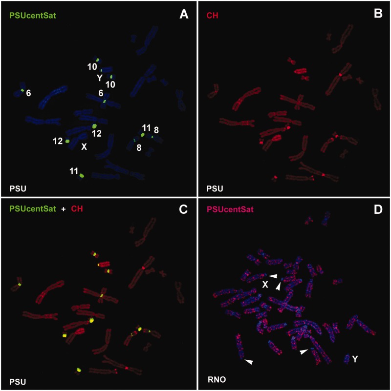
Fig. 2.—
Physical mapping of PSUcentSat on chromosomes of PSU and RNO. (A) Representative in situ hybridization presenting the chromosomal localization of PSUcentSat on chromosomes of PSU. The sequence was labeled with digoxigenin-11-dUTP and detected with 5′-TAMRA (red), but here it is presented in the pseudocolor green. Chromosomes were counterstained with DAPI (blue). (B) Same metaphase after sequential CBP-banding. Chromosomes were counterstained with propidium iodide (red). (C) Overlapping of PSUcentSat hybridization signals with C-bands. (D) Representative in situ hybridization presenting the chromosomal localization of PSUcentSat on chromosomes of RNO. Arrowheads evidence a depletion of PSUcentSat at the (peri)centromeric regions of some RNO chromosomes.

