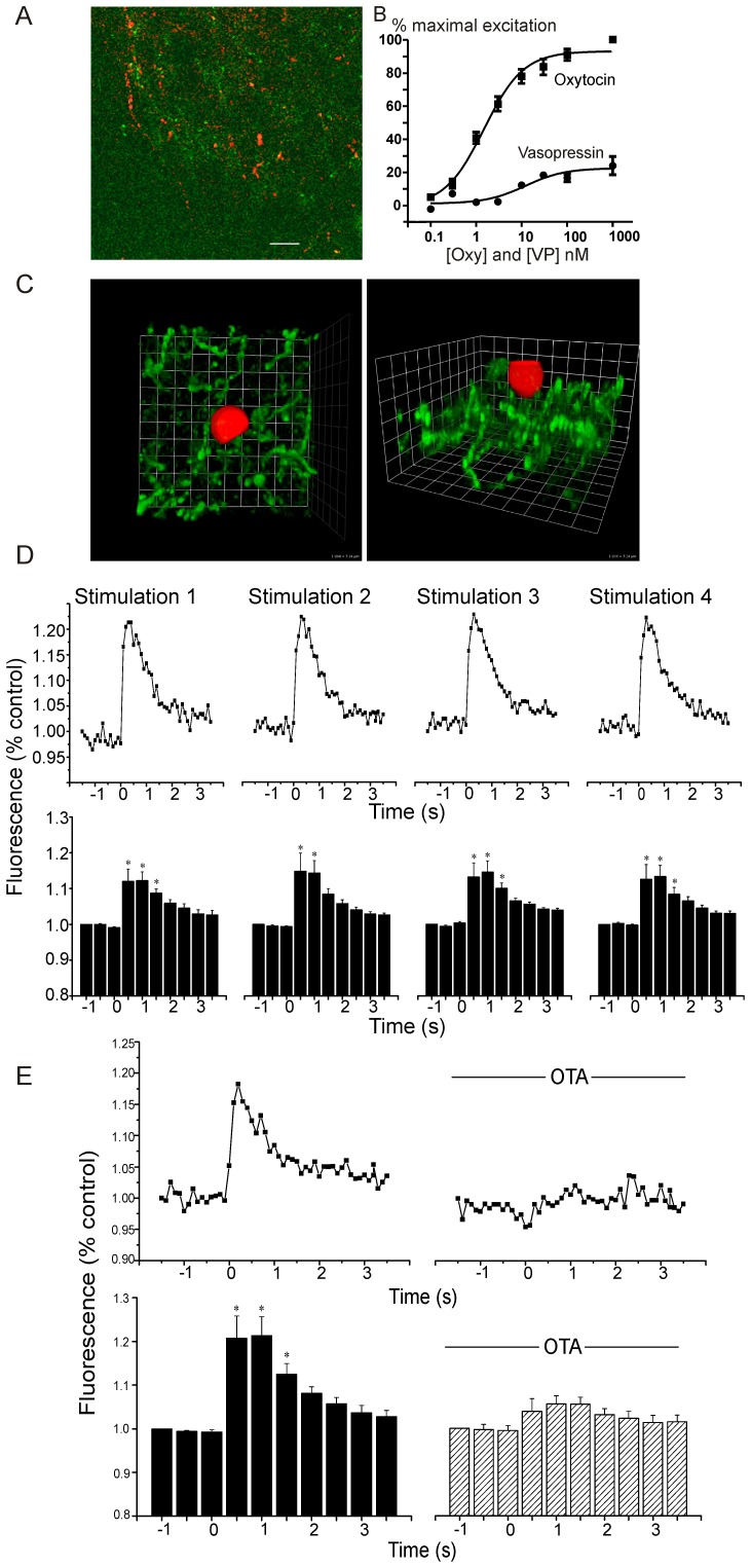
Figure 1. Imaging oxytocin and oxytocin receptor activation.
(A) Co-localization of ChR2-EYFP and oxytocin in brainstem fibers within the DMNV is shown (scale bar represents 28 microns). 55.7+3.7% of ChR2-EYFP PVN fibers in the DMNV are positive for oxytocin. (B) Both the oxytocin and vasopressin dose-response relationships of sniffer CHO cells expressing an oxytocin receptor and a R-GECO Ca2+ indicator were characterized. These oxytocin receptor expressing CHO cells are considerably more sensitive and responsive to oxytocin than vasopressin with a half maximal response (EC-50) for oxytocin of 1.5 nM, and an EC-50 for vasopressin of 12.1 nM. Responses to oxytocin were considerably more robust than that for vasopressin; at the concentration (1 µM) at which oxytocin maximally activates these cells, vasopressin evoked a blunted response of only 24+5% of the oxytocin response. Sniffer CHO cells deposited on slices with ChR2 PVN fibers (green) in dorsal motor nucleus of the vagus (DMNV) (C) detect optogenetic oxytocin receptor activation in brainstem DMNV tissue in close apposition to PVN fibers, (3-D reconstruction top down view (left) and side view (right)). D, Repeated stimulations (5 min apart) of ChR2 axons in DMNV increased Ca2+. Representative traces of one sniffer CHO cell, top, and averages of repeated stimulations in 9 CHO cells, * p<0.0001, bottom. E, Oxytocin antagonist OTA blocks Ca2+ response, representative trace of one cell, top, and average control increase in 7 cells (; * p<0.0001), bottom.

