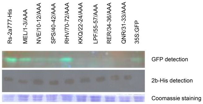
Figure 7. Immunoblot analyses of accumulation His-tagged 2b protein mutants in agroinfiltrated patches.
Detection of the fluorescence of GFP proteins on SDS-PAGE by illuminating the gel with UV lamp. A penta-his antibody was detection of His-tagged 2b proteins. Coomassie staining was used to monitor the equivalence of protein loading and transfer.

