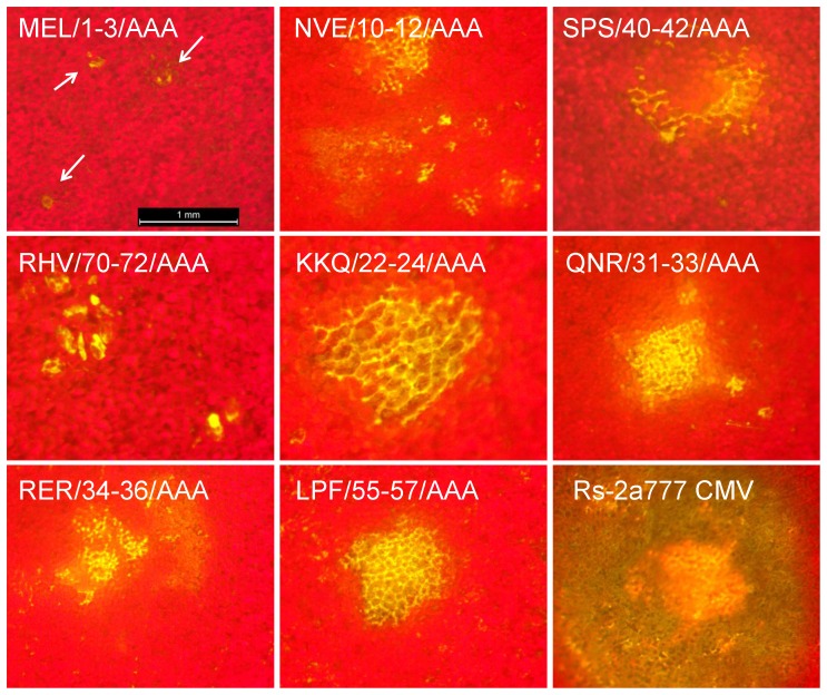
Figure 9. Development of fluorescence in Chenopodium murale plants infected with GPF-expressing derivatives of Rs-CMV and the eight mutant (MEL/1-3/AAA, NVE/10-12/AAA, SPS/40-42/AAA, RHV/70-72/AAA, KKQ/22-24/AAA, QNR/31-33/AAA, RER/34-36/AAA and Rs2LPF/55-57/AAA) constructs.
The red background is chlorophyll fluorescence from intact epidermal and mesophyll cells. Green-yellow represents the GFP-derived fluorescence from the chimera virus and the autofluorescence from necrotic tissue. The arrows point at the single epidermal GFP-fluorescent cells.

